Nociceptive pathways in the human brain handle the adventure of pain signals—from that first prick when you touch something sharp or hot to the moment your mind decides what that discomfort really means. Brain imaging studies have become super useful for checking out how these signals move and where things might go off track in chronic pain or sensory disorders. In this walkthrough, I’ll share how researchers use different brain imaging tools to dig into nociceptive pathways, what’s been learned so far, a few challenges, and some extra tidbits for folks curious about what’s really happening inside the brain when pain gets involved.
The Basics: What Are Nociceptive Pathways?
Nociceptive pathways are the networks that carry pain signals from your body up to your brain. When you experience something that might cause you harm, such as touching a hot stove, special sensors in your body called nociceptors fire off quick messages. These messages travel through your nerves, heading up the spinal cord and reaching the brain. Once there, the brain gets to work figuring out how much the pain bothers you and what you should do about it. Tracking down exactly how these pathways work is super important in pain research. It not only shows how pain starts and moves, but also sheds light on why certain types of pain feel the way they do.
Nociceptive pathways don’t just handle obvious injuries either. They play a role in migraines, certain gut issues, and even phantom limb pain. Researchers aim to unravel these pathways to develop better treatments and interventions. If you’re curious about how these sensors operate in different body parts, medical textbooks and trusted websites offer detailed diagrams and real-world cases to check out.
How Brain Imaging Sheds Light on Pain Processing
Brain imaging, or neuroimaging, uses technology to take detailed pictures of what’s happening inside the brain. When it comes to nociceptive pathways, these tools help show which regions “light up” during pain experiences, and how various conditions can mix things up in those patterns. The most popular types of brain imaging used for pain research include:
- fMRI (Functional Magnetic Resonance Imaging): Measures blood flow, offering a way to see which brain parts are busy processing pain when a stimulus is applied.
- PET (Positron Emission Tomography): Uses a small bit of radioactive tracer to track chemical changes. This lets scientists watch brain chemistry shift in real time as pain is experienced, which adds an extra layer of insight.
- EEG (Electroencephalography): Catches the brain’s electrical signals in fast bursts, letting researchers spot exactly when pain signals move from the body up to the cerebral cortex.
- MEG (Magnetoencephalography): Watches for the faint magnetic fields brain cells produce, so scientists can map how pain signals flow across brain areas.
Each method brings a new dimension of information. For example, fMRI and PET give a “where’s the action” snapshot, while EEG and MEG provide a “when did this happen” story. If you want to get more background on how these work, the National Institute of Neurological Disorders and Stroke offers an all-in-one summary of brain imaging methods worth reading.
What Brain Imaging Studies Have Shown about Nociceptive Pathways
Over years of research, brain imaging has helped map the highway system of pain and shown how our minds make sense of discomfort. Here are some highlights from these studies:
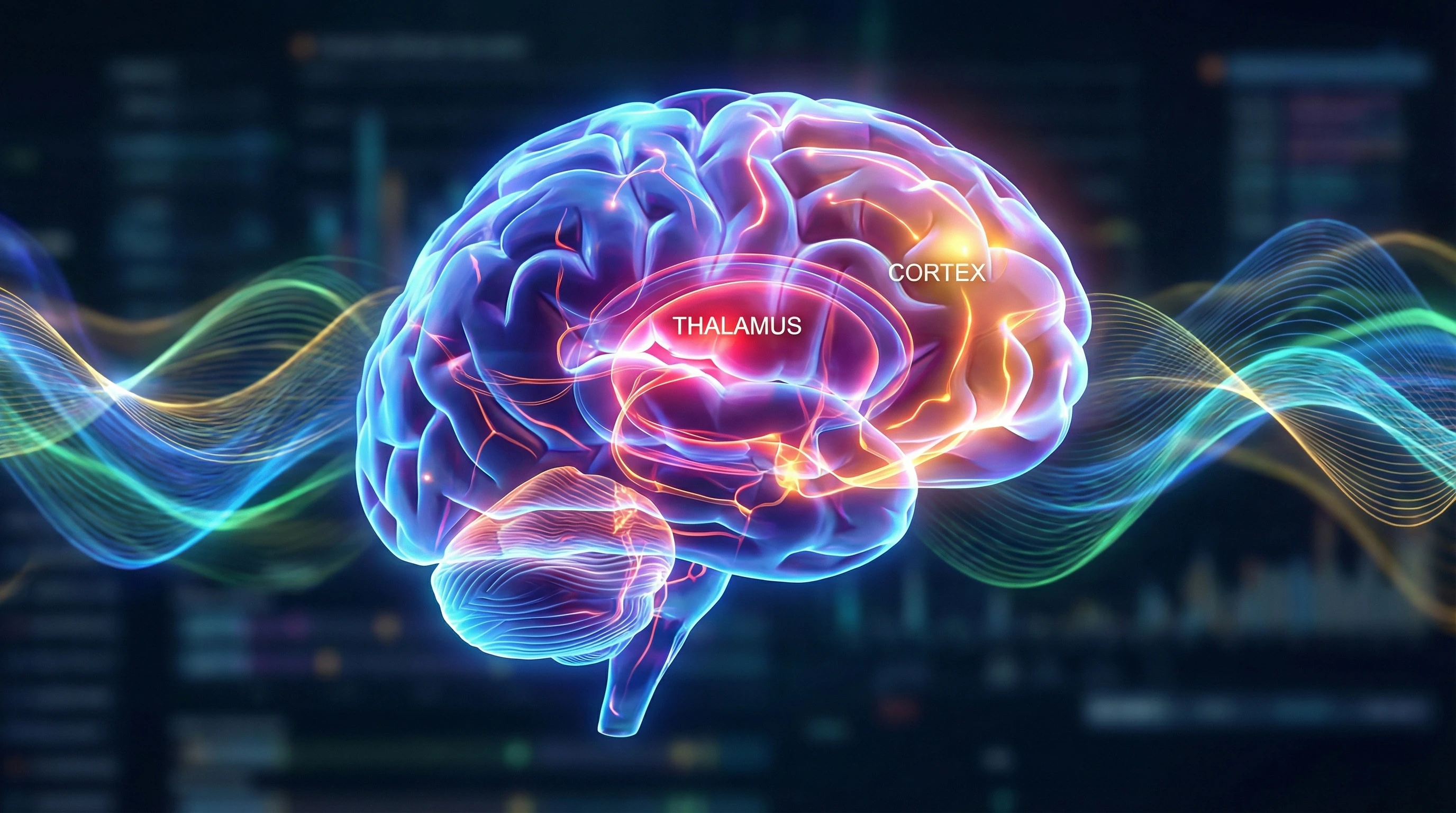
- Pain Matrix Activation: There’s a cluster of brain regions known as the “pain matrix”—including the thalamus (relay hub), somatosensory cortex (touch perception), insular cortex (emotions), and anterior cingulate cortex (attention/unpleasantness). These areas light up on scans during even mild pain. It’s not just about injury; even imagining pain can set some of these regions off.
- Expectation and Emotion: It turns out that expecting pain, or feeling anxious about it, stirs up the same parts of the pain matrix. Anxiety can make pain feel worse because your brain is already primed and alert, which explains why stress often ramps up pain experiences.
- Chronic Pain Changes the Brain: People living with chronic pain show a remixed network pattern: the pain matrix might stay more active or link up oddly with areas tied to memory and emotions. This often means pain feels stronger or sticks around longer, even after the original injury fades away.
- Modulation by Thoughts: You can actually give your pain perception a boost or dial it down. Cognitive tricks like distraction, meditation, or just picturing pain as less intense can lower activity in pain parts of the brain. Brain scans show this isn’t just mind over matter—it has a real, physical basis!
Diving deeper, studies using advanced techniques have started to show differences between acute pain (like stubbing your toe) and chronic pain conditions. Newer reviews are always being published, but a standout example is this NCBI review, which lists out key discoveries in the field of pain imaging.
Getting Started with Brain Imaging in Pain Research
If you want to get into this type of research, there’s a fairly standard process both for scientists and curious learners. Here’s how a typical brain imaging pain study unfolds:
- Select Volunteers or Patients: Recruit people—some healthy, some with chronic pain—to participate in the study.
- Set Up a Controlled Pain Stimulus: Use things like mild heat, safe electrical zaps, or pressure. The goal is to spark pain pathways without causing harm, always with ethics boards keeping a close watch.
- Acquire Scans During Stimuli: Volunteers’ brains get scanned during both pain and non-pain moments, so researchers can compare.
- Analyze Data for Activity Patterns: Cutting-edge software highlights which regions get busy. Comparing results across participants reveals trends or key differences.
- Draw Meaningful Conclusions: Is there overactivity in a specific cortex? Does mental distraction calm down brain responses? Are there signature differences in chronic pain?
Universities and hospitals with imaging tech often run these studies. If you’re thinking of getting involved, many of these places let students or the public volunteer to be scanned, offering a cool and educational experience.
Common Challenges and Practical Considerations
Studying pain in the brain isn’t easy. Researchers run into lots of everyday issues, including:
- Individual Differences: Some folks feel pain more sharply than others, so what stands out as “painful” in one scan may look mild in another. That’s why researchers need large, diverse groups to spot the real trends.
- Movement Artifacts: Scans need you to stay really still. Even tiny flinches or wiggles can mess up images, especially because pain can make people jump. Cushioned restraints and reminders to stay relaxed are pretty common tools.
- Pain’s Complexity: Emotional state, tiredness, and recent experiences all influence pain perception and brain activity. Experiments must juggle this complexity while making sure everyone is safe and comfortable.
- Resolution and Limits: Tech like fMRI offers clear spatial details but doesn’t zoom in on individual cells or moment-to-moment changes. Combining different scan methods takes extra time and resources, but provides the fullest picture available.
Movement Artifacts
Holding perfectly still in an MRI scanner can be surprisingly tough—especially if you’re waiting for something uncomfortable. Most labs use foam pads, flexible headrests, or even calming audio tracks. When a scan gets marred by too much movement, researchers often need to repeat it, frustrating both the teams and the participants. Some volunteers also share how the clanging sounds and close quarters add to the difficulty.
Complexity of Pain Experience
The feeling of pain is tied into memories, emotions, and even expectations. Researchers tackle this with detailed questionnaires, pain scales, and mental health assessments before the scan starts. They want a set of results that represents as much real life as possible, not just sterile lab pain.
In clinical practice, these complexities shape how chronic pain gets managed. Doctors may reference brain imaging to tailor both medication and therapies. Studies routinely explore how sleep, diet, and emotional support can shift pain outcomes, reminding us that pain never exists in isolation.
Deeper Insights: What Else Have We Learned?
Brain imaging continues to produce new and fascinating discoveries beyond simply mapping where pain happens. Some major insights include:
- Chronic Pain Biomarkers: New research points to certain brain activity patterns that may predict if someone’s pain will turn chronic. This could help catch and treat pain disorders early.
- Therapies Shaped by Imaging: Seeing how brain circuits change when people try medication, therapy, or meditation offers proof of benefit and clues for refining future treatments—for example, whether a technique lowers activity in pain-processing hubs.
- Pain Insights for Special Groups: Children, seniors, or people with neurological differences process pain in unique ways. With imaging, care teams can customize support, ensuring no group is left out.
In practice, researchers use fMRI to check how pain medicine affects real brain circuits, digging into how both the drug and someone’s attitude toward it can help lower pain. These methods now extend into trials for non-opioid drugs and innovative therapies, providing new paths away from traditional painkillers.
Frequently Asked Questions
Folks interested in pain imaging research often want to know:
How safe are brain imaging studies involving pain?
Answer: These studies use only mild and carefully supervised pain stimuli. Imaging methods like fMRI and EEG have no radiation risk; PET involves a trace amount of radioactivity. All studies require strict ethical approval and thorough safety checks.
Can imaging tell if someone is “faking” pain?
Answer: While imaging can’t “read minds,” it does show real brain patterns that are hard to fake. Still, pain is personal and subjective, so scans help spot possible signatures of true or chronic pain but can’t act as lie detectors.
Is it possible to “read” pain in real time?
Answer: EEG and MEG are fast enough to catch brain signals as pain arrives, although figuring out the intensity takes more complex analysis. Using several imaging tools together gives a better sense of what’s going on overall.
Key Takeaways for Anyone Curious about Pain Research
Stumbling upon new discoveries about nociceptive pathways through brain imaging is helping scientists, doctors, and patients all get a clearer picture of what happens when pain hits the brain. New funding and better tech are helping studies get more all-in-one and precise, boosting the chance for treatments that fit how pain is really processed in our heads. Staying curious—whether by volunteering in a study, reading the newest research, or just keeping an eye out for fresh tools—is a great way to stay connected if you’re passionate about neuroscience or health care.
Learning how the brain processes pain through these images doesn’t just satisfy curiosity. It’s making real differences for patient care, mental health support, and how we treat pain moving forward. Science is always rolling forward, sharpening the picture a bit more every year—which means better relief may be closer than we think.