Pain is a complex experience involving not just the body but how the brain interprets hurt. To really get a sense of how pain is processed, researchers tap into neuroimaging techniques to check out what goes on inside the brain during painful situations. These technologies let us visualize brain activity, spot the regions involved in pain, and even compare how different people or conditions may mix up pain responses. In this article, I’ll walk you through some of the most asked-about neuroimaging techniques used to study pain perception in the brain, with clear explanations along the way.
Why Study Pain Perception with Neuroimaging?
Pain is not just about injury or physical harm. It is also shaped by emotions, memories, and even what we expect might happen. If I feel pain from a stubbed toe, someone else’s reaction to that same injury could be totally different. This variety makes studying pain in the brain extremely important. Neuroimaging methods help me track down which brain areas light up when people face different types of pain, from heat on the skin to headaches or ongoing back discomfort. These studies shed light on not just why pain varies from person to person, but also how pain management and treatments can get better.
Research in this area has grown fast, especially over the last few decades. It has shifted the understanding of pain from only being a physical problem to being a process rooted in the brain. Seeing these changes in real brain activity gives doctors and scientists the tools to develop more effective ways to diagnose, treat, and even prevent chronic pain.
Neuroimaging Basics: Main Tools for Pain Research
There are several key neuroimaging techniques used to study pain. Each comes with its own strengths in catching what is going on in the brain, either in real time or over longer periods. Here are the most popular tools:
- Functional Magnetic Resonance Imaging (fMRI): This technique follows changes in blood flow in the brain. When a part of my brain works harder, it uses more oxygen. fMRI spots these changes and lets researchers map out where pain is handled. Since it is noninvasive and shows details clearly, it is a top pick for pain research.
- Positron Emission Tomography (PET): PET scans use bits of radioactive tracers to measure how the brain uses energy. These tracers highlight active regions, so scientists can see how pain is handled and what brain chemicals are released during a pain experience.
- Electroencephalography (EEG): EEG uses sensors on the scalp to catch the brain’s electrical activity. It excels at showing quick shifts in brain signals, making it perfect for spotting what happens just as pain hits. While it cannot pinpoint exactly where in the brain the signal comes from, it’s good at timing.
- Magnetoencephalography (MEG): MEG picks up the magnetic fields created by the brain’s electrical currents. It is fast at reading timing, like EEG, but usually gives a better sense of the general location where pain shows up in the brain.
Sometimes, researchers mix these methods to get a broader picture. One tool might be great for location details, while another works better for speed, so using them together can really give a boost to the results.
How Neuroimaging Sheds Light on Pain Perception
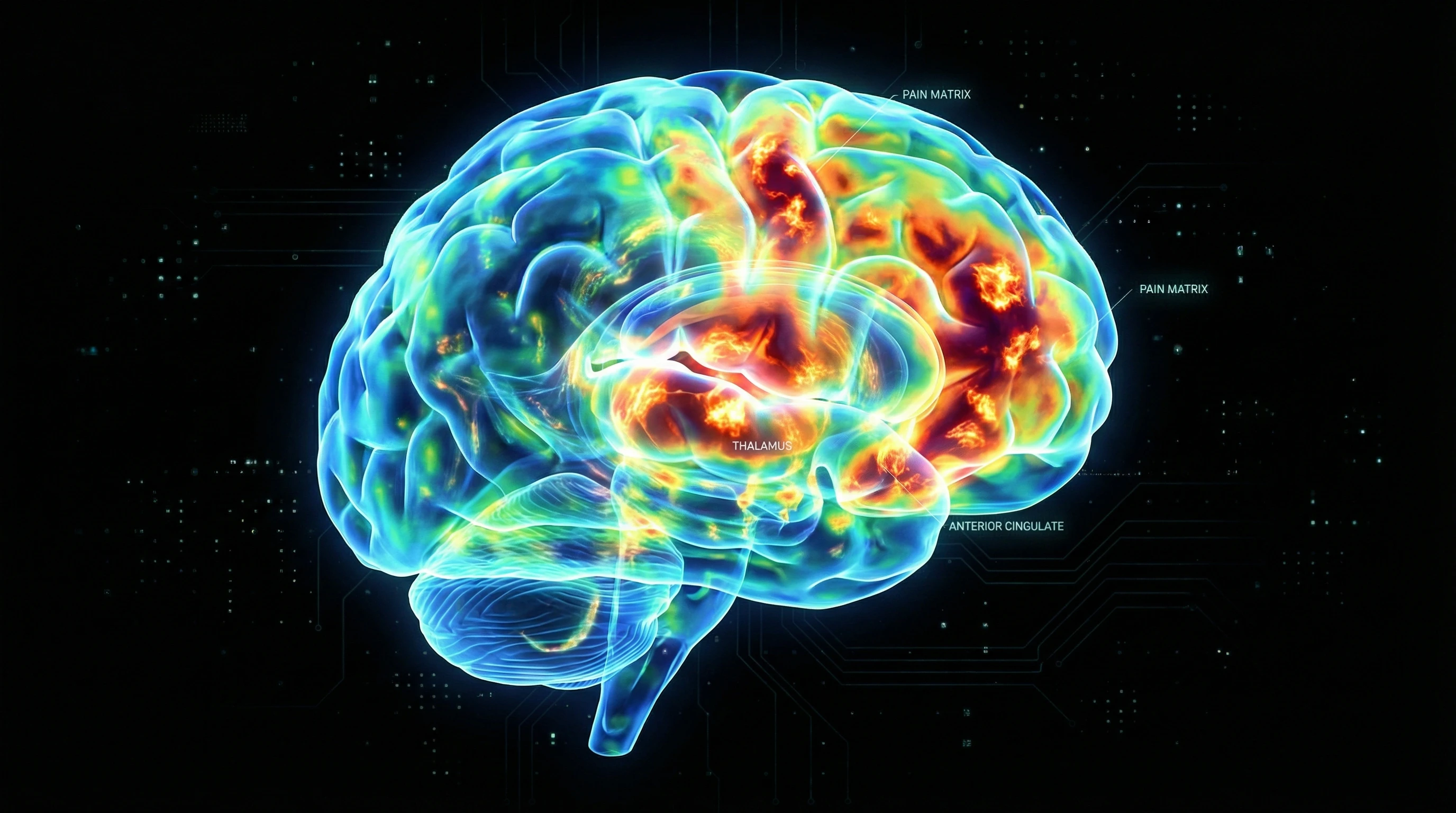
When I look over research on brain imaging and pain, I see certain regions light up again and again. These areas, often called the “pain matrix,” include the somatosensory cortex (handling the physical side), the insula (tying physical states with emotions), the anterior cingulate cortex (linked with how we feel pain), and the thalamus (which relays signals in the brain).
Researchers ask lots of creative questions using neuroimaging in pain science, such as:
- How does pain from different causes—like heat, cold, or pressure—look inside the brain?
- What changes for people who end up with chronic pain after an injury, compared to those whose pain gets better?
- How do treatments like medications, meditation, or even therapy change brain activity connected with pain?
- Why do some people feel pain much more strongly than others, even when the trigger is the same?
By giving a once-over to these patterns across people and pain types, scientists start to make sense of the blend of body and mind that shapes each person’s pain.
Essential Concepts to Understand Before Exploring Neuroimaging Research
Jumping into neuroimaging research can seem technical, but having some basic ideas on your radar makes it easier. Here are great starting points:
- Temporal vs. Spatial Resolution: Temporal resolution means how quickly a tool can pick up changes in the brain. EEG and MEG are extremely fast, working in milliseconds. Spatial resolution is about how clearly a technique maps out brain locations—fMRI and PET shine in this department.
- Invasiveness: Most current neuroimaging methods for pain are noninvasive, so there’s no surgery needed. PET does use a safe, tiny dose of radioactive stuff.
- Data Interpretation: Scans show changes tied with pain, but not pain itself. Scientists need to carefully compare the scans with self-reports or behavior for accurate conclusions.
Knowing these ideas helps if you start reading pain science journals, work in labs, or just want to see how cutting-edge treatments are developed.
Common Challenges and Smart Solutions in Pain Neuroimaging
Despite all the cool things neuroimaging can do, there are still some challenges. Here’s what I often see, and smart ways researchers are handling them:
- Variability in Pain Experience: Everyone feels pain a little differently. To tackle this, studies ask people to rate their pain or use larger groups to spot trends that stand up in many cases.
- Motion Artifacts: Moving during a scan, even if it’s just a bit, can mess up results. Since being in pain might make you fidget, researchers use careful instructions, shorter scans, and improved equipment to tone down this issue.
- Placebo Effects: Just believing a treatment will work can change the brain’s pain signals. Science teams use placebos and control groups to catch and account for this when they break down the data.
- Cost and Accessibility: Since imaging gear is pricey, studies might have smaller samples or only focus on specific pain types. People team up with hospitals and universities to pool resources and get more out of their data.
With careful planning and fresh technology, these hurdles are being lowered as the field grows.
Real-World Impact: How Neuroimaging Changes Pain Treatment
The lessons learned from neuroimaging are actively shaping how pain is treated. For example, clinical trials for new drugs may use imaging not just to check if the pain matrix settles down, but also to see if parts tied to emotion or memory react differently. In therapy like cognitive-behavioral interventions for chronic pain, brain scans sometimes show shifts in both pain-related and emotion-linked regions after progress is made.
Doctors also use these scans to figure out tricky pain disorders that don’t show up in regular tests or when symptoms don’t make sense with obvious injuries. Looking ahead, neuroimaging could help customize treatments, sending patients toward the approach that sparks the best brain response for their pain type.
- Personalized Pain Management: Advanced brain imaging may let doctors pick which medicines or therapies will have the highest chance of working, especially if a person’s pain is tough to treat.
- Preventing Chronic Pain: Catching unusual brain patterns early after injury could help stop pain from becoming long-lasting, jumping in with support before patterns get locked in.
Real examples like these show that understanding pain in the brain is not just about science studies. It directly helps people get better pain care and live more comfortably.

Frequently Asked Questions
Here are a few questions I often hear about neuroimaging and pain:
Question: How do neuroimaging scans show pain if pain is a personal feeling?
Answer: Scans can’t show pain directly, but they do show changes in brain activity that line up with people’s own descriptions of pain. By putting together what patients say and what the scans reveal, scientists get closer to understanding pain in the brain.
Question: Can brain imaging diagnose chronic pain?
Answer: Scans help rule out other conditions and show differences between people with and without ongoing pain, but a single scan usually isn’t enough for a clear diagnosis. Doctors mix imaging, medical exams, and history to sort things out.
Question: Is it safe to get a neuroimaging scan for pain research?
Answer: Yes, most scan procedures—like fMRI, EEG, and MEG—are safe for the majority of people. PET scans do involve a very small dose of radiation, so they’re used carefully and only when necessary.
Wrapping Up
Neuroimaging has transformed what we know about how the brain reads pain. These techniques act as a window into both the physical and emotional sides of discomfort, helping connect the dots between people’s symptoms and actual brain changes. It’s truly fascinating seeing these breakthroughs make pain care more personal, precise, and tuned in to what’s really happening in the brain. The future looks bright for tracking down even smarter ways to prevent and treat pain, all thanks to what neuroimaging continues to put out there.